ფაილი:Computed tomography of human brain - large.png

თავდაპირველი ფაილი ((3 639 × 2 595 პიქსელი, ფაილის ზომა: 3,9 მბ, MIME ტიპი: image/png))
| ეს ფაილი მდებარეობს Wikimedia Commons სერვერზე. იხილეთ მისი აღწერის გვერდი სრული ინფორმაციისთვის. |
 |
გადასვლა ფაილის გვერდზე |
| ეს ფაილი ვრცელდება Creative Commons CC0 1.0 Universal Public Domain Dedication ლიცენზიით. | |
| პიროვნებებმა, რომლებიც ერთობლივად მუშაობდნენ ამ ნამუშევარზე, გადაწყვიტეს ამ ნამუშევრის გადაცემა საზოგადოებრივ საკუთრებაში, უარი განაცხადეს ნამუშევრის ყველა უფლებაზე მსოფლიო საავტორო უფლებების კანონის იმ ფარგლებში (მათ შორის სხვა უფლებებიც) რომელიც დაშვებულია კანონის მიხედვით. თქვენ შეგიძლიათავტორის ნებართვის გარეშე დააკოპიროთ, შეცვალოთ, გაავრცელოთ, გამოიყენოთ აღნიშნული ნამუშევარი ნებისმიერი მიზნით, მათ შორის კომერციულითაც.
|
|

| აღწერა |
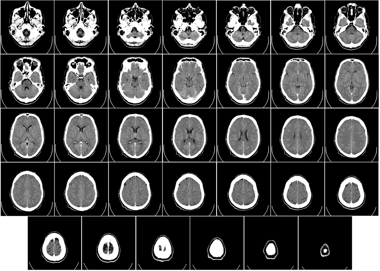
English: Computer tomography of human brain, from base of the skull to top. Taken with intravenous contrast medium.
It was taken Mars 23, 2007 on the uploader, after a 20 minute episode of homonymous hemianopsia with loss of the left visual field, but nothing strange was found. Three episodes of scotoma occurred in the following years, whereof the last one was scintillating (depiction). Otherwise, there were no further neurological symptoms.
Türkçe: Geçirdiği bir kaza neticesinde homonim hemianopsi vakası oluşan bir hastanın beyninin bilgisayarlı tomografisi. Tomografi neticesinde bir anomaliye rastlanmamıştır. |
| თარიღი | Uploaded January 17, 2008 |
| წყარო | Radiology, Uppsala University Hospital. Uploaded by Mikael Häggström. |
| ავტორი | Department of Radiology, Uppsala University Hospital. Uploaded by Mikael Häggström. |
| უფლება (ფაილის მეორეული გამოყენება) |
Compound images
-

-
 Inverted


Scrollable stack
For larger version, see Category:Computed tomography images of Mikael Häggström's brain. To move through the images, hover over the image and use scroll wheel, drag the mouse, or click the < or the > above each stack. This functionality should activate when the page is fully loaded, which may take some time.

.png)
.png)
.png)
.png)
.png)
.png)
.png)
.png)
.png)
.png)
.png)
.png)
.png)
.png)
.png)
.png)
.png)
.png)
.png)
.png)
.png)
.png)
.png)
.png)
.png)
.png)
.png)
.png)
.png)
.png)
.png)
.png)
.png)
.png)
Case with multiplanar reconstruction
-
 Brain, case 1: Multiplanar, but no intravenous contrast.
Individual images
Licencing
| ეს ფაილი ვრცელდება Creative Commons CC0 1.0 Universal Public Domain Dedication ლიცენზიით. | |
| პიროვნებებმა, რომლებიც ერთობლივად მუშაობდნენ ამ ნამუშევარზე, გადაწყვიტეს ამ ნამუშევრის გადაცემა საზოგადოებრივ საკუთრებაში, უარი განაცხადეს ნამუშევრის ყველა უფლებაზე მსოფლიო საავტორო უფლებების კანონის იმ ფარგლებში (მათ შორის სხვა უფლებებიც) რომელიც დაშვებულია კანონის მიხედვით. თქვენ შეგიძლიათავტორის ნებართვის გარეშე დააკოპიროთ, შეცვალოთ, გაავრცელოთ, გამოიყენოთ აღნიშნული ნამუშევარი ნებისმიერი მიზნით, მათ შორის კომერციულითაც.
|
DICOM format
ფაილის ისტორია
დააწკაპუნეთ თარიღზე/დროზე ფაილის დასათვალიერებლად, როგორც ის მაშინ გამოიყურებოდა.
| თარიღი/დრო | მინიატიურა | ზომები | მომხმარებელი | შენიშვნა | |
|---|---|---|---|---|---|
| მიმდინარე | 01:11, 24 დეკემბერი 2017 | | 3 639×2 595 (3,9 მბ) | Shashi. | Reverted to version as of 12:49, 1 February 2008 (UTC) |
| 10:59, 8 მაისი 2008 |  | 3 639×2 595 (3,17 მბ) | CountingPine | Optimise using PNGOUT | |
| 12:49, 1 თებერვალი 2008 |  | 3 639×2 595 (3,9 მბ) | Mikael Häggström | {{34 computer tomography images}} {{Individual images of CT of Mikael Häggström's brain}} | |
| 11:56, 31 იანვარი 2008 |  | 3 639×2 595 (4,03 მბ) | Mikael Häggström | {{34 computer tomography images}} {{Individual images of CT of Mikael Häggström's brain}} |
ბმულები
ამ ფაილზე ბმული მოცემულია შემდეგ გვერდებზე:
ფაილის გლობალური გამოყენება
ეს ფაილი გამოიყენება შემდეგ ვიკებში:
- გამოყენება bn.wikipedia.org-ში
- გამოყენება bo.wikipedia.org-ში
- გამოყენება ca.wikipedia.org-ში
- გამოყენება en.wikipedia.org-ში
- CT scan
- Portal:Medicine
- Portal:Medicine/Selected picture
- Portal:Medicine/Selected picture archive
- Wikipedia:WikiProject Neuroscience
- Wikipedia:Featured pictures/Sciences/Biology
- User:Mikael Häggström
- User talk:Mikael Häggström/Archive 1
- Wikipedia:Featured pictures thumbs/10
- Wikipedia:Featured picture candidates/CT of brain of Mikael Häggström.png
- Wikipedia:Featured picture candidates/February-2008
- Wikipedia:Wikipedia Signpost/2008-02-11/Features and admins
- Portal:Medicine/Selected picture/9, 2008
- Portal:Medicine/Selected picture/9
- Wikipedia:Picture of the day/July 2008
- Template:POTD/2008-07-11
- Wikipedia:Wikipedia Signpost/2008-02-11/SPV
- User:Mikael Häggström/Gallery
- Wikipedia:WikiProject Medicine/Recognized content
- Computed tomography of the head
- Wikipedia:Wikipedia Signpost/2013-10-02/Op-ed
- Wikipedia:Wikipedia Signpost/Single/2013-10-02
- User:Wouterstomp/test
- User:Fitness queen04/sandbox
- Wikipedia:WikiProject Anatomy/Resources
- Wikipedia:WikiProject Anatomy/Recognized content
- Wikipedia talk:WikiProject Anatomy/Archive 9
- Reconstruction from projections
- User:VGrigas (WMF)/Quality Media
- User:Flyer22 Frozen/Human brain
- Portal:Medicine/Recognized content
- User talk:Rhododendrites/Reconsidering FPC on the English Wikipedia
- გამოყენება es.wikipedia.org-ში
- გამოყენება fi.wikipedia.org-ში
- გამოყენება he.wikipedia.org-ში
- გამოყენება hy.wikipedia.org-ში
- გამოყენება hyw.wikipedia.org-ში
- გამოყენება id.wikipedia.org-ში
- გამოყენება is.wikipedia.org-ში
- გამოყენება ja.wikipedia.org-ში
იხილეთ, ამ ფაილის გლობალური გამოყენება.